VIDEOLAPAROSCOPIC SURGERY USING THE ROBOT (ROBOTIC SURGERY)

Videolaparoscopic surgery using a robotic platform, commonly called Robotic Surgery, emerged in recent years as the last frontier of technical development applied to videosurgery. As such, it is nothing more than robot-assisted video-surgery.

Robotics adds some advantages to the purely laparoscopic method. With the robotic system, the surgeon has a real three-dimensional view of the surgical field and its positioning, seated with arms supported, provides excellent ergonomics.

Robotic surgery instruments were specially developed to give the surgeon a range of motion similar to that of the human hand and can achieve rotation of up to 360 degrees.

The freedom of movement of robotic tweezers provides fast and intuitive learning. The robot can also be adjusted to perform movements with reduced scale amplitude, which allows sutures in small spaces with great aplomb. In addition, it eliminates tremors giving great precision of movements.

INFORMAÇÕES DO AUTOR:

Dr. Marcel Autran Machado Especialista em cirurgia do aparelho digestivo e cirurgia minimamente invasiva

Professor livre-docente de Cirurgia pela Universidade de São Paulo, graduado pela Faculdade de Medicina da USP (FMUSP) e com residência em Cirurgia Geral e do Aparelho Digestivo pela mesma instituição.
CRM-SP nº 70330

In advanced laparoscopic procedures, there is often a need for staplers to control large vessels. The robot allows sutures very quickly and quickly, which makes it possible to control large vessels without the use of endoscopic staplers.

The initial disadvantage of this method was the cost, which was gradually reduced with the greater use of the method and partnerships between doctors and hospitals. Today, some hospitals do not charge any extra fees for using the robot in some complex procedures, such as liver, pancreas and biliary tract surgeries.

This method was implemented in Brazil and Latin America in early 2008. That same year, we began our experience with pioneering robotic liver¹ and pancreas² surgeries (published at the time).

In simpler videolaparoscopic procedures, such as cholecystectomy and uncomplicated appendectomies, robotic surgery adds little to the advantage, since these procedures are very easily performed by the conventional videolaparoscopic method. However, we have routinely used the robot in some less complex procedures such as correction of gastroesophageal reflux, correction of inguinal hernias and other hernias of the abdominal wall.

In simpler videolaparoscopic procedures, such as cholecystectomy and uncomplicated appendectomies, robotic surgery adds little to the advantage, since these procedures are very easily performed by the conventional videolaparoscopic method. However, we have routinely used the robot in some less complex procedures such as correction of gastroesophageal reflux, correction of inguinal hernias and other hernias of the abdominal wall.

The biggest advantage in using robotic surgery occurs in the most complex procedures. In the field of general surgery and the digestive tract, the procedure s that benefit most from this method are pancreas, liver and biliary tract surgeries.

Since 2018, we have been using the robot in all these surgeries instead of laparoscopy. With this, we were able to increase the proportion of patients who benefit from the minimally invasive method, because in some situations where the use of laparoscopy was not possible and we needed the use of open surgery, today with the robot this is not only possible but also desired.

An example of this is its use in Klatskin³ tumors, that is, hilar cholangiocarcinoma³. Another important example is its use in pancreatic cancer. Surgeries such as pancreaticoduodenectomy4 or Whipple surgery4 are routinely performed by our team robotically today. In cases with vascular invasion, previously a contraindication regarding the use of laparoscopy, the use of the robot facilitates its safe performance in selected cases.

Another frequent use of the robot occurs in patients who have already undergone multiple open surgeries, where most surgeons in the world contraindicate minimally invasive surgery due to the presence of adhesions. Robotic surgery helps a lot in the release of adhesions and thus, we are able to perform surgeries, even complex ones, in these patients.

The use of the robot in this situation will provide a better postoperative result, as well as allow further surgeries to continue to be performed by a minimally invasive route, as once adhesions are released using this technique, they do not form again with the same intensity. A liver surgery5 in this extreme situation was the subject of a publication in an international journal5.

Another important use is in complex bile duct surgeries such as in cases with bile duct stenosis (narrowing) caused by accidental injuries during previous surgeries or by other causes. A bile duct surgery in this extreme situation was the subject of one of our publications in an international journal6.

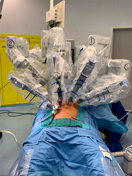
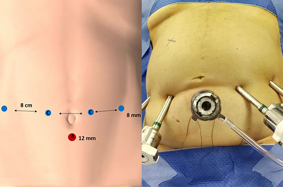
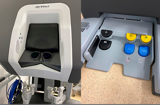
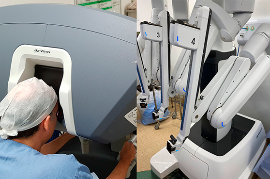
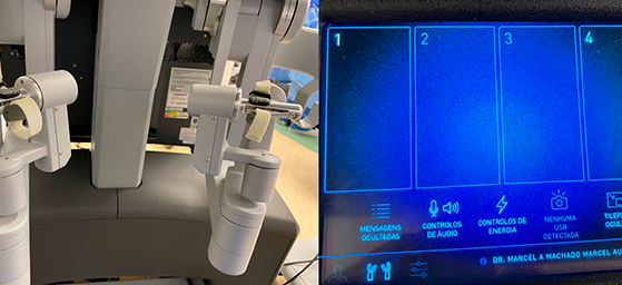
How is robotic surgery performed? Through small incisions (Image 1), through which the trocars enter (cannulas through which the different robotic forceps, optics and other instruments enter). The robot uses 4 trocars. An extra trocar is used by the assistant surgeon to introduce sutures, aspirator, stapler, etc. (Image 1). The surgeon is on a console (Image 2) from where he commands the movement of the 4 robot arms (camera and 3 tweezers). (Images 3 and 4). Video 1 shows how the robotic system works.

  • 1 – 063 Robotic liver resection. Report of pioneering experience in Latin America
  • 2 – 135 Robotic Resection of Intraductal Neoplasm of the Pancreas
  • 3 – 215 Robotic Resection of Hilar Cholangiocarcinoma
  • 4 – 200 Robotic pancreaticoduodenectomy after Roux-en-Y gastric bypass
  • 5 – 198 Robotic Repeat Right Hepatectomy for Recurrent Colorectal Liver Metastasis
  • 6 – 203 Robotic Left Hepatectomy and Roux-en-Y Hepaticojejunostomy

IMAGE 1

The schematic drawing shows the type and position of the trocars used for robotic surgery. Camera (C) is used in two different positions. The auxiliary port (12 mm) used by the bedside surgeon (red sphere). A minimum distance of 8 cm must be maintained between the robotic arms (blue spheres).

IMAGE 2

On the left we see the console with 3-D view. On the right we see the control pedals. On the console the surgeon is able to control the position of the camera and three robotic arms, two at the same time and the third is positioned for assistance and is fixed. To do this, the surgeon uses a combination of pedals and arms (handles).

IMAGE 3

On the left, we see the main surgeon sitting at the console with a 3-D immersion view during the procedure. On the right, we see the robot before being covered with a sterile cover and introduced into the surgical field (docking).

IMAGE 4

On the left we see the manual controls (handles) closely. On the right, we see some of the controls available to the surgeon during the procedure.

VÍDEOS

1 - 3 de 27